Free Shipping for all Order of ₹500
COD payment is available.


How Bacterial Cell Walls Decide the Final Colour in Gram Staining
How Bacterial Cell Walls Decide the Final Colour in Gram Staining
A stained bacterial smear can reveal an unexpected contrast under the microscope. Some are a deep purple shade and others appear pink. The contrast raises an important question: why do bacterial cells respond differently to the same stains?
Gram staining answers that question with a series of carefully controlled steps. The method separates two main groups based on differences in the bacterial cell walls. It is a mixture of several reagents, each having a special part to play. The last colour provides information on cell behaviour during the staining process.
What Is Gram Staining?
Gram Staining method is used to differentiate bacteria according to their cell wall structure. It classifies bacteria into Gram positive and Gram negative.
The method begins by adding a purple primary stain to the bacterial cells. Then iodine helps the cells hold the stain in the next stages. The major difference between the two groups is the decolorization. A counterstain is then used to give a contrasting colour to cells that lost the primary stain.
This technique converts a microscopic smear into a clearly visible pattern. The result does not simply show that bacteria exist. It also gives an early indication of their structural group.
What Causes the Colour Difference?
The principle of Gram staining is based mainly on differences of the bacterial cell envelope. Gram-positive bacteria are characterised by a thick layer of peptidoglycan.
Crystal Violet first enters the bacterial cells and gives them a purple colour. Gram’s Iodine then interacts with Crystal Violet and forms a larger stain complex.
The decolourizer creates the critical separation. The thick peptidoglycan layer in Gram-positive cells helps to retain the stain complex. The decolourizer also causes the cell wall to become less permeable.

Gram-negative cells respond differently during this stage. Their outer membrane allows the decolourizer to remove lipids from the cell envelope. The thinner layer of peptidoglycan does not retain the stain complex as well.
Then the Safranine enters the decolourized cells and stains them pink or red. The primary stain is still dominant so the gram-positive cells are still purple.
From Smear to Microscope
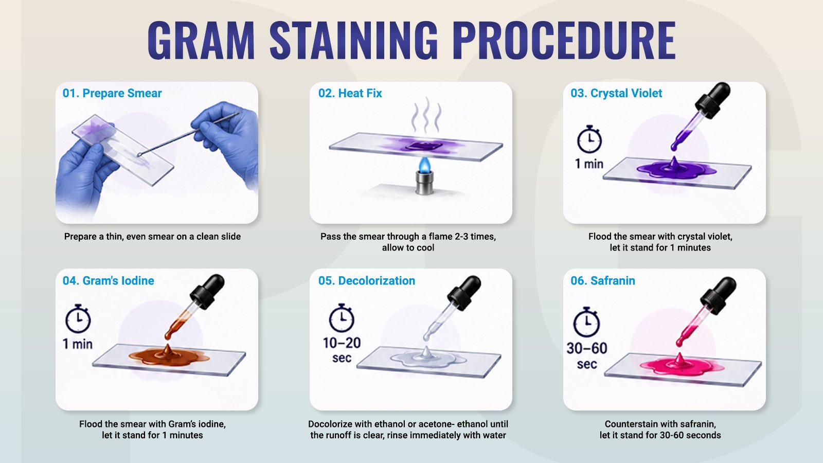
A proper gram staining procedure begins with a clean bacterial smear. The smear should dry before heat fixation. Heat fixation helps attach the bacterial cells to the glass slide.
Next, apply Crystal Violet to cover the smear. Allow the stain to act before rinsing the slide gently with water.
Apply Gram’s Iodine over the smear. This step is required for the stain complex formation inside the bacterial cells.
Decolourization must be carried out carefully. Briefly apply the decolourizer and watch carefully for runoff. Too much decolourization can have an impact on the final result.
After decolourization, apply Safranine as the counterstain. Rinse the slide and allow it to dry before microscopic examination.
Gram’s Stain - Kit (TK 004) brings the four main reagents together in one kit. It includes Crystal Violet, Iodine, Decolourizer, and Safranine. This arrangement supports the complete staining sequence with the required reagents.
Why Decolourization Matters
The principle of gram staining is best understood during decolourization. Before this stage, both bacterial groups carry the purple stain complex. The decolourizer is responsible for the visual distinction that later can be seen under the microscope.
Gram-positive cells have a thick layer of peptidoglycan which helps them to keep the complex and retain the stain. The loss of the complex from the Gram-negative cells is due to different interaction of the decolourizer with the cell envelope.
This phase needs attention, as the timing affects the final interpretation. Too much decolourizer can remove more stain than expected. Too little can give Gram-negative cells a false purple appearance.
Reading the Final Result
The final colour provides the basic result.
Gram-positive bacteria stain purple or violet because they retain the primary stain. Gram negative bacteria lose the primary stain and pick up the counter stain Safranine and appear red or pink.
The observer then notes the shape, arrangement and Gram reaction of the cells. These observations are useful for further identification of bacteria.
Examples of Gram-Positive and Gram-Negative Bacteria
Several familiar bacteria show these two Gram reactions.
Gram-positive examples include Staphylococcus, Streptococcus, and Bacillus. Gram-negative bacteria include Escherichia coli, Salmonella, and Pseudomonas.
These examples help connect the final colours with common bacterial groups seen in microbiology.
Conclusion
A smear that is stained can tell more than a field of tiny cells. The colour pattern shows differences in bacterial cell structure.
Gram staining uses a series of stains, iodine, decolourizer and counterstain, to make those differences visible. Each stage is described so that the final purple and pink pattern is more easily interpreted during routine microscopic examination.
To explore the Gram’s Stain - Kit (TK 004) and learn more about it, click here.
Frequently Asked Question (FAQs)
Q1. Can Gram staining identify a bacterial species?
A. No. Gram staining provides a preliminary classification depending on staining reaction, shape and arrangement. More tests are needed to determine the species.
Q2. Why should fresh bacterial cultures be used?
A. In older cultures, the staining reactions may not be uniform. Fresh cultures usually give more reliable and better Gram reactions.
Q3. Why is heat fixation used?
A. It is helpful to firmly attach the bacterial cells to the slide.
Q4. What happens if the smear is too thick?
A. With a thick smear the decolourisation may be uneven. It could also make it hard to see individual cells clearly under a microscope.
Q5. What are Gram-variable bacteria?
A. Gram-variable bacteria show both purple and pink cells in the same preparation. Such appearance may be due to several factors, including age of culture and staining conditions.

leave a reply
Your email address will not be published. Required fields are marked *





The information below is required for social login
Sign In
Create New Account